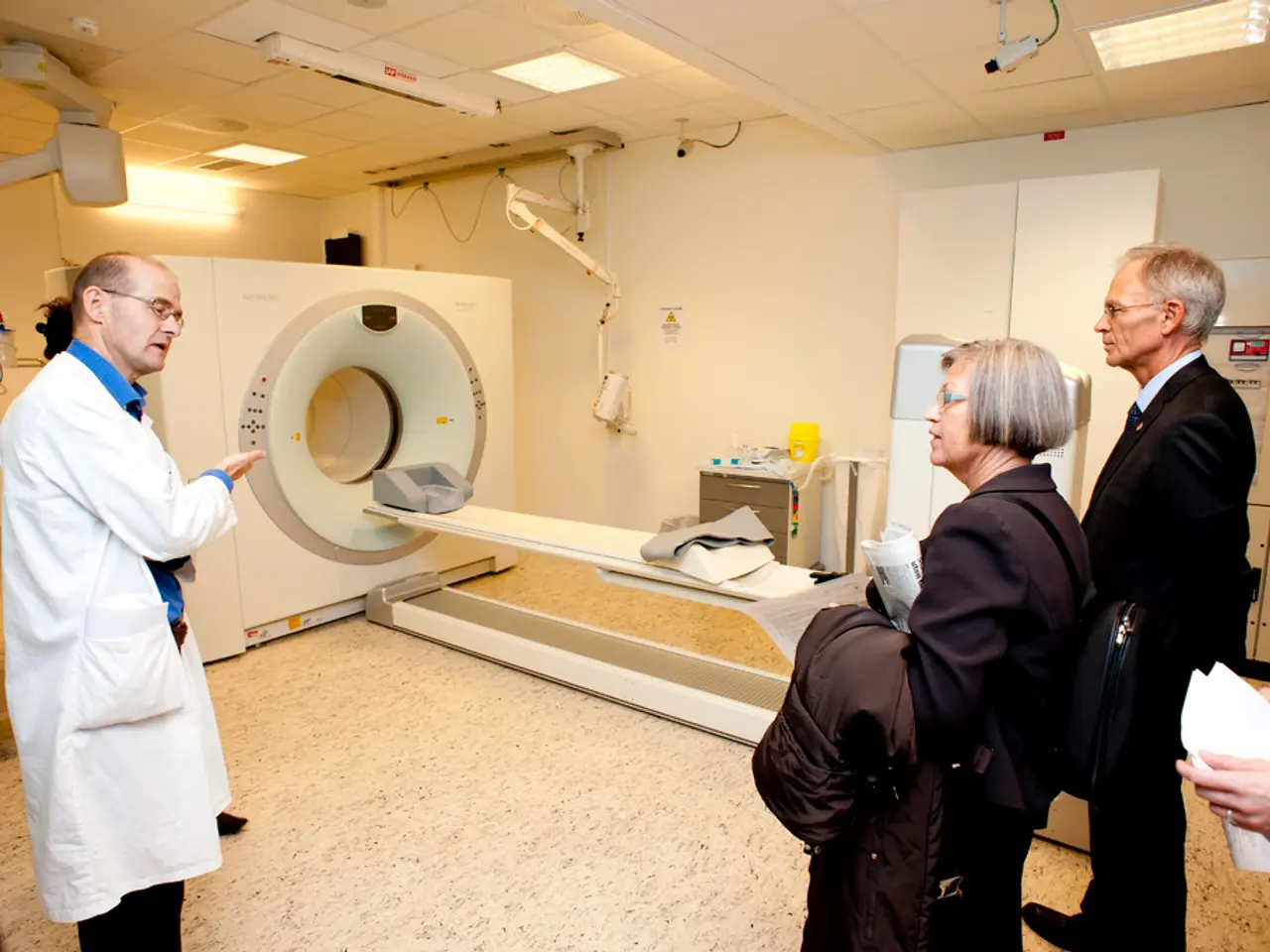
Scan for Heart Perfusion Imaging: Preparation, Potential Risks, and Step-by-Step Procedure
A heart perfusion imaging scan is a non-invasive diagnostic procedure that assesses blood flow to the heart muscle, playing a critical role in both diagnosing ischemic heart disease and tailoring patient-specific management strategies.
This scan is particularly valuable in detecting obstructive coronary artery disease by showing regions of the heart with inadequate blood supply during stress (exercise or pharmacologic) compared to rest. It also helps evaluate the functional significance of coronary artery blockages and differentiate between viable heart muscle and scar tissue from prior heart attacks.
Perfusion scans can guide clinical decision-making on the need for interventions like angioplasty or bypass surgery. They are also useful in assessing recovery or progression of myocardial perfusion abnormalities over time in patients undergoing treatment.
Heart perfusion imaging can be performed using various modalities such as cardiac MRI, CT, or nuclear techniques. Stress perfusion imaging, which simulates exercise conditions pharmacologically, reveals ischemic areas by detecting perfusion defects not visible at rest.
In management, perfusion imaging helps identify patients at high risk due to significant ischemia who may benefit from revascularization. It also monitors myocardial viability and scarring to inform prognosis, evaluates right ventricular function, and tracks improvement or deterioration after treatment.
Doctors may recommend periodic heart perfusion scans for people with known heart conditions or those at risk of heart disease to monitor heart function and identify changes or progression in heart conditions. After the scan, most people can resume normal activities immediately, but if the test involves a stress component, people may feel tired and require rest.
However, there is a risk of radiation exposure associated with heart perfusion imaging scans, and doctors discuss this risk with patients before the test. It is best to drink plenty of water after the scan to flush the radiation from the body.
Before the scan, people should avoid caffeine and nicotine and not eat anything for at least 4 hours. People with severe asthma or chronic obstructive pulmonary disease (COPD) should inform their doctor before getting a heart perfusion imaging scan, as some may experience side effects or complications from the medication and exercise during a stress test.
During a resting scan, a small amount of a radioactive tracer is injected into a vein in the person's arm, and images are taken using a gamma camera. If stress testing is involved, the person may walk on a treadmill or use an exercise bicycle while images are captured, or doctors may administer medications to stimulate the heart.
People should discuss their current medications with their doctor, who may recommend stopping medications such as beta-blockers or calcium channel blockers before the test. Ischemia, which can cause reduced heart strength, can be determined by a heart perfusion imaging scan. A healthcare team will closely monitor the person's vital signs during the scan.
Chest pain, especially during physical activity or times of stress, can prompt doctors to recommend a heart perfusion imaging scan. After assessing the distribution of the radioactive tracer in the heart, a cardiologist will interpret the images obtained from the scan and identify areas of reduced blood flow or abnormalities.
In conclusion, heart perfusion scans provide detailed anatomic and functional information about myocardial blood flow and viability, aiding in the diagnosis and management of heart disease.
- Heart perfusion imaging scans are crucial in detecting and diagnosing obstructive coronary artery disease, particularly during stress conditions, which may lead to patient-specific management strategies.
- In addition to assessing coronary artery disease, these scans can evaluate mental health, cardiovascular health, and fitness and exercise levels by identifying regions of the heart with inadequate blood supply.
- These non-invasive scans are beneficial in the health-and-wellness sphere, as they help monitor and manage medical-conditions such as myocardial perfusion abnormalities over time.
- In the sports domain, heart perfusion imaging scans can be used to identify athletes at risk due to significant ischemia, who may require revascularization to ensure cardiovascular health.
- Similarly, sports-betting enthusiasts might find this information valuable for making informed decisions on the potential health and performance risks of their chosen athletes.


